ADNEXAL MASSES & OVARIAN CYSTS
What are Adnexal Masses or Ovarian Cysts?
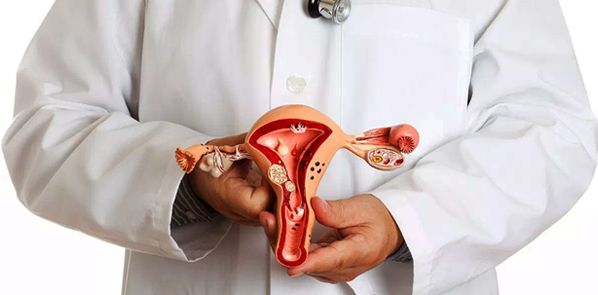
The pelvic organs in the abdominal cavity are the uterus, fallopian tubes and ovaries along with its blood vessels and support structures. Adnexa refers to the area connecting to the uterus, such as the fallopian tubes and ovaries. When a mass occurs in this region, it is referred to an adnexal mass.
Most adnexal masses develop in the ovary. Women of all ages can develop adnexal masses and may be discovered due to symptoms or incidentally from a physical exam, radiologic imaging or surgery. These masses can be cancerous or non-cancerous, therefore a workup should be performed to identify if these masses may be malignant or benign.

Some causes of adnexal masses are listed below:
Functional Cysts – During ovulation an egg is matured in a follicle that develops and then ruptures to release the egg. This signals a corpus luteum to grow to help maintain hormones if pregnancy is achieved. The corpus luteum gets resorbed if a pregnancy is not conceived. If the follicle does not rupture, it can continue to grow into a follicular cyst. If the corpus luteum does not get resorbed and continues to grow, it is called a corpus luteal cyst.
Polycystic Ovary – This ovary is enlarged due to the development of many small follicles. Typically, this is seen in women who have polycystic ovarian syndrome (polycystic ovaries, infrequent or lack of menses, and/or evidence of high male hormones).
Endometrioma – Ovarian cyst that contains tissue from the uterine lining or endometrium; also referred to as a “chocolate cyst” because the fluid inside is old blood produced from the endometrial tissue in the cyst and looks like chocolate. This develops as a process of endometriosis.
Dermoid – (aka Mature Cystic Teratoma) This cyst arises in the ovary and is a benign tumor consisting most commonly of hair, fat and teeth. This cyst is common in women between the ages of 20 and 40. If less well-differentiated tissues such as brain, bone, or glands are present then this mass is considered malignant (Immature Teratoma).
Fibroma – This is a solid benign tumor of the ovary that may be associated with fluid in the abdomen and lungs (Meigs’ Syndrome). This is usually seen in postmenopausal women.
Cystadenoma – This is a common benign tumor that can contain serous or mucinous fluid within the cyst.
Other benign tumors of the ovary – There are other tumors of the ovary that are benign, but some may produce increased levels of different types of hormones, such as estrogen (Granulosa cell tumor), androgens or male hormones (Sertoli-Leydig cell tumors) or thyroid hormone (Struma Ovarii).
Tuboovarian Abscess – (TOA) This is a collection of pus in the tubes and ovaries from PID (pelvic inflammatory disease) that is usually accompanied with symptoms of abdominal pain, fever and vaginal discharge. PID is sexually transmitted and can cause infertility. The tuboovarian abscess implies acute infection and therefore requires immediate attention.
Ectopic Pregnancy – This is when a pregnancy forms outside of the uterus. Most commonly an ectopic pregnancy is in the fallopian tube and may cause pain. If you have a positive pregnancy test and a sudden onset of pelvic pain, please call your physician immediately because these pregnancies can outgrow the fallopian tube, rupture and cause severe bleeding.
Hydrosalpinx – A benign process of fluid getting trapped inside a fallopian tube. This may cause pain and decrease fertility rates.
Cancer – Cancer may develop in the ovary or fallopian tube. Other cancers, especially from breast and the gastrointestinal tract, may spread to the adnexal region as well.
Fibroid – This is a benign tumor of the uterine muscle that may grow adjacent to the uterus, presenting itself in the adnexal region. See website link Fibroids.
If the ovarian mass is large, ovarian torsion can occur. This is defined as the ovary twisted upon its own blood supply. Ovarian torsion may completely cut off the blood supply resulting in a nonfunctional or “dead” ovary. Any type of adnexal mass, benign or malignant, can undergo torsion. Typically, a woman with torsion presents with pelvic pain, possible low grade fever and an adnexal mass.
How are adnexal masses diagnosed?
The first important step is discussing your medical history with your physician. A family history is also important in determining if you at an increased risk for cancer. Your physician will also ask you about symptoms you may or may not be experiencing to help with the diagnosis, such as abnormal uterine bleeding, weight change, and pain.
Next, your physician will perform a physical exam to identify signs of disease. Your physician may be able to palpate the mass and identify from which structure it arises from as well as describe its size, tenderness, consistency, mobility and if it is well-defined or not. The exam is helpful in narrowing down the list of causes so that you do not have to undergo any unnecessary labs or imaging.
One of the most valuable imaging studies we use to identify and characterize adnexal masses is the ultrasound. Adnexal masses on ultrasound have certain characteristics to help your physician identify what type of a mass it is. Blood flow to the ovary can also be visualized on ultrasound, which can help with the diagnosis. Other imaging, such as a CT scan or MRI, may be useful as well.
The main concern with adnexal masses is whether or not they are malignant. A tumor marker called CA-125 is ordered if suspicion for ovarian cancer is high. This tumor marker is not used as a screening tool for ovarian cancer because it can be elevated with many other conditions, such as endometriosis, PID, and fibroids. CA-125 is most useful to follow the levels in those patients already diagnosed with ovarian cancer. There are other tumor markers for different types of ovarian cancers and based on your history, your physician may order some of these as well.
Another important lab test is a pregnancy test to rule out an ectopic pregnancy. Other lab tests may be ordered by your physician to help with making a diagnosis, such as androgen hormone levels to help with the diagnosis of polycystic ovarian syndrome.
How are adnexal masses treated?
Once the diagnostic workup for the adnexal mass is completed, your physician may discuss with you conservative, medical or surgical management. The management options will be based on your age, medical history, physical exam and lab and imaging workup.
Below is some treatment options of the more common adnexal masses:
Functional Cysts – Observation is appropriate with serial ultrasounds to make sure that these cysts are not growing or developing concerning features. Some women are just “cyst formers” and birth control pills may be suggested to prevent ovulation and the formation of these cysts. Surgery is recommended to preserve the ovary if the cyst is large enough to undergo torsion.
Polycystic Ovary – There are no specific treatments for the ovary itself, however women with the syndrome may consider weight loss and birth control pills to help regulate their menstrual cycles. If pregnancy is desired, ovulation induction medications can be used.
Endometrioma – These cysts usually do not spontaneously regress or respond to medication. Therefore surgery may be recommended for removal.
Dermoid – Surgery is recommended to prevent growth, torsion, and rupture.
Fibroma – Since this cyst is prevalent more in postmenopausal women, surgical removal of that ovary and tube is recommended. Preservation of the ovary can also be performed with removal of the fibroma only.
Cystadenoma – Surgical removal is recommended to prevent growth, torsion, rupture, and rule out malignancy.
Other benign tumors of the ovary – Surgical removal is recommended to prevent growth, torsion, rupture, and rule out malignancy.
Tuboovarian Abscess – Hospital admission is required to monitor for signs of the infection spreading to the bloodstream and response to intravenous (IV) antibiotics. Drainage of the abscess may be performed, especially if symptoms do not improve within 24-48 hours. Abscess drainage can be performed under radiologic imaging guidance or can be surgically excised.
Ectopic Pregnancy – In an asymptomatic patient a medication, called Methotrexate, can be administered in the office with serial ultrasound and lab follow-up until the pregnancy is resorbed. Depending on the severity of the symptoms or characteristic of the ectopic pregnancy, the patient may be a candidate for medical or surgical therapy.
Hydrosalpinx – If fertility is desired, the tube may need to be surgically repaired or removed. Without fertility desire or symptoms, conservative management may be appropriate.
Cancer – A referral to an oncologist is strongly recommended for a thorough discussion of the management.
Fibroid – Please see website link Fibroids.
If the workup for an ovarian mass is thought to be malignant, a referral to a gynecologic oncologist is strongly recommended. If the ovarian mass is thought to be benign, then preservation of the ovary is strongly recommended with only removing the cyst (cystectomy) from the ovary. On occasion the entire ovary may be removed. If this is performed, generally the fallopian tube is removed as well. This procedure is called a Salpingo-oophorectomy. Any specimen, either the cyst wall or the ovary, is sent to pathology to confirm whether it is benign or malignant. We perform our surgeries through the laparoscope as an outpatient surgery for quicker recovery and less pain. Please see link Laparoscopy.
Our goal at the Advanced IVF Institute and the Advanced Gynecologic Surgery institute is to provide the best care possible. Please fill out the form below to request a consultation with us.
